Looking at tissue from up close
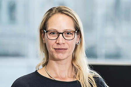
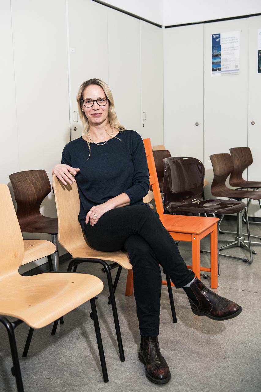
Dr. Kühl, you sit at a microscope every day and look at cells. What’s the source of your motivation to do this work?
What’s long fascinated me in research is that you don’t google an idea or a question, but instead answer it yourself. Initially, I studied Biotechnology and treated the waste water from paper bleaching works. But when you’re busy carrying 20-litre waste pails and it smells bad, at some point you start wishing that you’re back in the immunology lab – that’s how I got into medical research. Apart from that, I think it’s important to see what you’re researching for. That’s why I find the structure at the Benjamin Franklin Campus practical: here, we have two ward blocks where the patients are treated and, between them, the research department – which means there’s close contact between doctors and scientists. The doctors see what the patients need and can in turn communicate it to the researchers. I think this idea has been well implemented here in this building – you can't come up with new ideas on your own.
You’re the main user of a device that Professor Siegmund requested within the framework of the BIH Investment Fund. What kind of device is it?
In histopathology, we examine tissue sections under the microscope according to color. In the iPATH.Berlin we offer an elaborate method for this purpose, where it is not worthwhile for everyone to perform it in their own laboratory. We therefore have clients not only within the Charité, but also Berlin-wide and internationally. With conventional transmitted light microscopy, you can look at samples that are for example marked with color precipitates in red, brown and blue. Or you use a fluorescence microscope and look at samples that have been made to shine by means of fluorescent colorants. However, nowadays there are flow cytometers that measure up to 30 different parameters at the single cell level. So, histopathology, with its three or four colors, can hardly keep pace with that. When you color several markers on one tissue section, in the end you can hardly differentiate between them with the naked eye. The new microscope has a camera that takes multispectral pictures. That means that even though one sees red or brown, the camera always recognizes where there’s a mountain and a valley. It can thereby differentiate between colors that the eye can’t tell apart from one another. This enables us to analyze several parameters on one tissue section, and thus makes us more attractive for scientists who otherwise only work with the flow cytometer.
And what’s the purpose of it all?
You can identify immune cells in the body based on different markers and thus create an immunological map. For many procedures, immune cells have to be dissolved out of the tissue and you get cells in the test tube: it’s no longer possible to say where those cells were located in the organ – however, that’s important for understanding a disease. For example, in the intestine there‘s a special layer of cells that separates the immune system of the intestine from its inner workings, including bacteria and food components. Here, there are cells that arrange themselves exactly underneath that cell layer. They identify components from the intestine and signal to the immune cells that these belong to the intestinal flora and are not supposed to trigger a reaction. It’s also important to be able to demarcate that cell layer, which is only possible in the tissue section. Conversely, it’s possible to look at a single cell and see what that specific cell is doing, what neurotransmitters it’s sending out and what state it’s in. We therefore try to also make it histopathologically possible to not only localize the cell concerned but also to observe its function.

Funding program
BIH Investment Fund
Funding period
2015
Project title
Immunohistochemical and immunofluorescent optical multiplexing for in situ characterization of functional cell populations
Research areas
Histopathology, gastroenterology
Institution
Charité – Universitätsmedizin Berlin
Since 2017
Head of the iPATH.Berlin – Core Unit Immunopathology for Experimental Models, Charité – Universitätsmedizin Berlin
2016
Teaching qualification in Experimental Medicine
2001 –2016
Research Fellow in, after 2011 head of, histopathological services of the Collaborative Research Centres SFB633 and SFB650, Charité – Universitätsmedizin Berlin
Yet looking at cells through a microscope probably wasn’t your childhood dream?
As a doctoral student, I studied chronic inflammatory bowel diseases and we always gave our tissue samples to a pathologist. He was able to explain things incredibly well and was enthusiastic about histopathology. So, I was not only able to have the results brought to me but also to look at it through the microscope myself and have it explained to me. He worked all day long for the clinic and nevertheless took time for an interested doctoral student. Later on, as a Professor at the Charité, Mr. Loddenkemper thought of me when he was building up his research team. Since then, that enthusiasm has never left me. I find it incredibly exciting to look at tissue sections through the microscope. I just find it nicer to see how thick a cell is, what it has for runners and where it sits than to see dots flashing in the flow cytometer.
What words of wisdom would you like to impart to younger scientists to help them on their way?
Young researchers should trust themselves. If they have any ideas that they want to pursue, they shouldn’t let themselves be held back or constrained by their teachers and professors. They often tackle a question in a more naive and inquisitive way and should be allowed to keep that quality for a bit longer.
December 2017 / MM
