In search of new methods: How multiple sclerosis can be detected at an early stage through an ultrasound scan of the eye
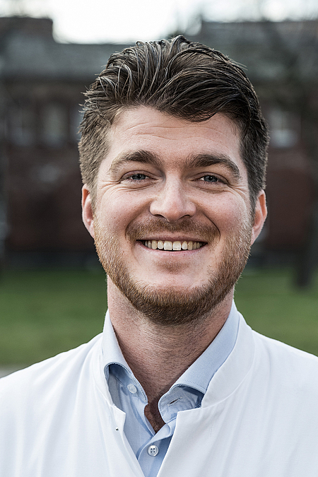
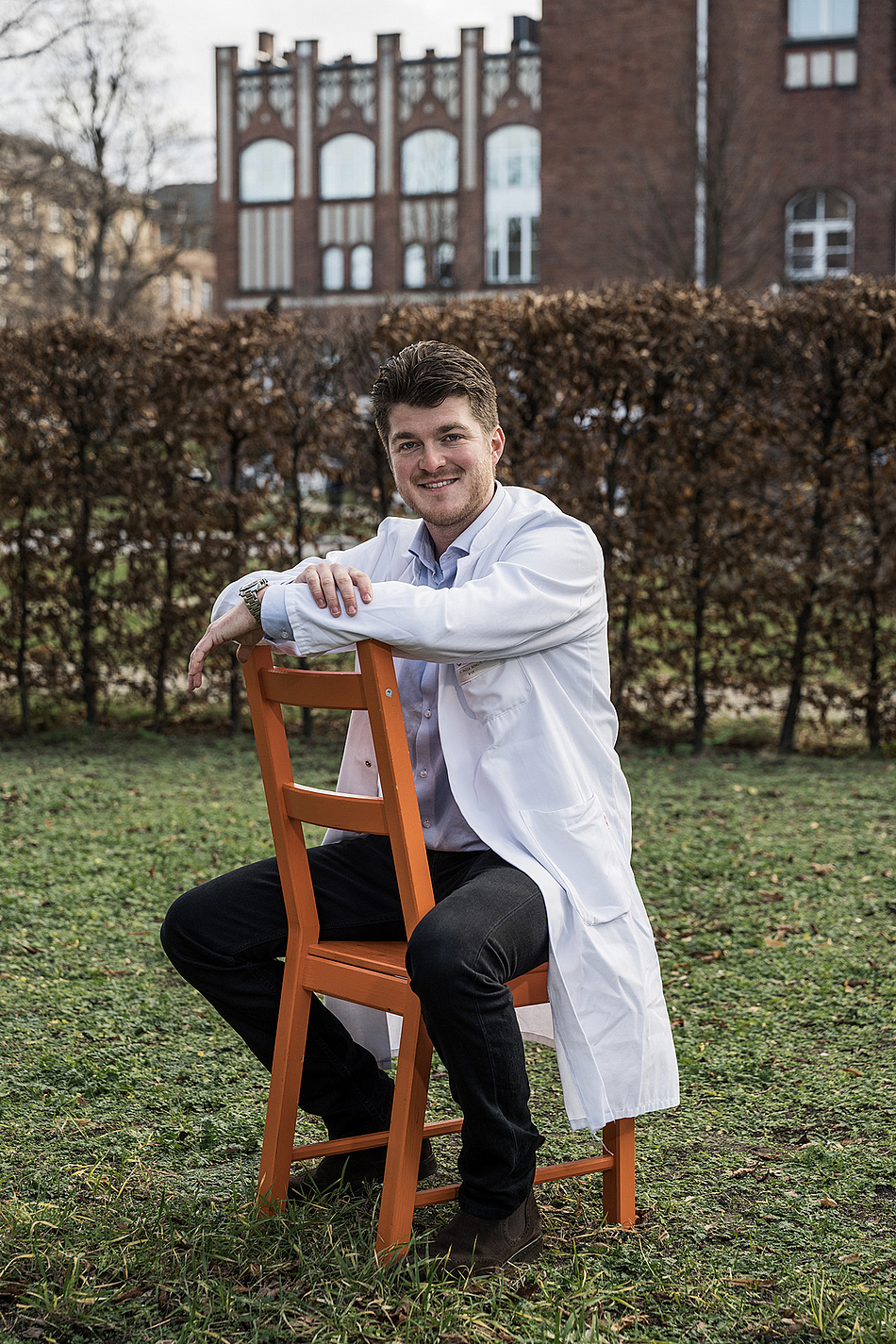
Dr. Felix Schmidt is a neurologist and specialist in multiple sclerosis. As a Clinician Scientist, he’s granted “protected time” to enable him to focus on his research project in addition to his work in patient care. He is testing a completely new method of diagnosing multiple sclerosis in a faster and simpler way – with an ultrasound scan of the eye. We talked with him about how the two things are connected, about the difference between clinical and scientific work and – not least – about his passion for travel and mountaineering.
Dr. Schmidt, what has an ultrasound scan of the eye got to do with multiple sclerosis (MS)?
MS is a chronic, inflammatory disease of the central nervous system. The most common first symptom among patients is an inflammation of the optic nerve, i.e. optic neuritis. 30 percent have it as their first symptom and a total of 60 percent of MS patients have one at some point in the course of the disease. This already shows the connection between MS and the eye. On the other hand, diagnosing optic neuritis is no trivial matter. This is the source of the dictum “The patient sees nothing and the doctor sees nothing.” One important clinical sign of optic neuritis is a so-called “RAPD”, a relative afferent pupillary defect. Here, the pupil light reflex is damaged. The pupil doesn’t contract when a light is shone into the eye, and sometimes even expands. This makes it sound as if it’s simple and reliable to detect, but that’s only true of the very pronounced cases. In milder cases, misinterpretations can occur – and this is where the ultrasound method comes into play.
And can you describe how an ultrasound scan of the eye is carried out?
The patient lies down on the examination table with their eyes closed. Then the ultrasound probe is held against their lower eyelid and, using a torch, you shine light through their closed eyes. The light is nevertheless perceived by the eye and the pupil reacts. It’s as simple as any other ultrasound scan. The pupil is measured when at rest, as well as under the influence of direct and indirect light. We measure the pupil’s diameter and how fast the pupil contracts under the influence of light. To begin with, we successfully tried it out on healthy patients and published norm values. In the case of patients with RAPD, these values are pathologically altered. Our study is the first ever to examine pupillary function using ultrasound, and also the first study to examine that of MS patients with an RAPD. At the moment, it’s still only patients from the Charité, but in future we hope to be able to include patients from outside the Charité in the study.
Is it possible to measure pupil function in other ways too? What are the advantages of your ultrasound method?
Yes, there are other methods of measuring it. The gold standard is measurement with a special camera, the pupillometer. This is a special examination and, in the field of practice-based medicine, it’s predominantly only ophthalmologists who have such devices. However, pupil function is also simply estimated visually. The ultrasound method has a number of advantages. It’s more precise, because you can measure the millimeters precisely. This makes the method more objectifiable. You can also save the results and document the process. So, it’s not just a method for improving diagnosis, but can also be used for assessing the effects of therapy. In addition, ultrasound devices are much more common than pupillometers. So, our method is more easily accessible and applicable in the broader clinical field. It’s even conceivable that it could have further areas of application too. The fact that the eye doesn’t have to be open for the examination, but is closed, offers many possibilities – e.g. in examining coma patients or patients with traumatic brain injuries whose eyes are swollen closed.

Funding program
BIH Charité Clinician Scientists
Funding period
2018 – 2021
Project title
B-Mode ultrasound as a non-invasive imaging biomarker for assessing the afferent visual system
Research area
Neuroscience
Institution
Charité – Universitätsmedizin Berlin
Since 2013
Assistant doctor at the Clinic for Neurology and member of staff at the University Outpatient Department for MS, Charité - Universitätsmedizin Berlin
2016
Guest scientist, Center for Sleep & Circadian Biology (Prof. P. Zee), Ken & Ruth Davee Department of Neurology, Northwestern University, Feinberg School of Medicine, Chicago, USA
2015 – 2016
Study physician and research assistant at NeuroCure, AG Neuroimmunology
What in fact led you to research MS?
Already during my studies, I always found the nervous system fascinating. I’m currently training to be a neurologist. I first studied MS intensively while writing my doctor’s thesis. The deeper I went into it, the more interesting I found the disease. The pathomechanisms are still unknown. There’s some very interesting research on the subject and many advances have been made in treatment options over the past few years. 20 or 30 years ago, it was certain that MS sufferers would eventually end up in a wheelchair. Today, we’re making steady progress in halting the progressive deterioration for the patients, i.e. stopping the activity of the disease. I find it extremely exciting to be working at the cutting edge of research in this field. I’ve had some excellent mentors who inspired my interest in neurology and MS and who were, and are, role models for me at both a professional and personal level. About four years ago, I was also offered the opportunity to assist with the MS consultation hour, and since then I’ve enjoyed doing it once a week. What I particularly like in my day-to-day work is that the patients are usually somewhat younger. With MS, the peak period for contracting the disease is from the age of 20 to 45.
As a BIH Clinician Scientist, you regularly alternate between clinical and scientific work. Do you regard yourself as more of a clinician or a scientist?
To be honest, I see myself as being exactly in the middle. It’s precisely the combination of the two that I like and I don’t want to miss either aspect. I really like the contact with patients. Also, at the clinic, you can see the effects on patients more quickly.
In research, you need a great deal of patience. It can take months or years until you discover something or get results. What motivates me in my research is the desire to find solutions that are of clinical relevance and benefit for the patients. Even after I complete my specialist medical training, I want to continue to combine both fields of activity.
Did you already want to be a doctor as a child?
No, when I was a child I wanted to be a pilot. My love of travel has remained, only nowadays I fulfil it differently. I completed some portions of my studies in France and Switzerland and did a research stay in the US.
Do you do a lot of private travelling too?
I really enjoy travelling and being in nature. I like going on ski tours and mountaineering trips. It’s my way of unwinding from work. A couple of months ago, I went on a three-week expedition to Island Peak in the Himalayas – at an altitude of 6,200 meters – with my father. It was unforgettable and I’m already looking forward to the next tour.
A playful question in conclusion: Which three people, living or dead, would you like to have an imaginary dinner with? I’m guessing you’d definitely pick Reinhold Messner?
Actually, that’s right – Reinhold Messner, and also Albert Einstein and Jimi Hendrix. It would probably be a hilarious dinner.
June 2020 / Marie Hoffmann
