Improved Patient Welfare in Radiology – The Vision for a Start-Up
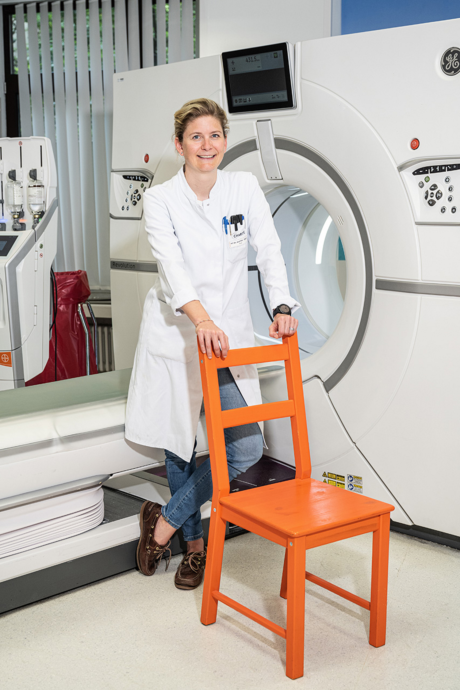
Katharina Erb-Eigner is originally from Austria and is one of the select few to have secured funding from the Inventors for Health (I4H) pilot program as part of Stiftung Charité’s focus on innovation. The funding from Stiftung Charité has enabled her to make great strides with RadioEye, an entrepreneurial project which joined the Digital Health Accelerator Program at the Berlin Institute of Health in 2022. We spoke to Dr Erb-Eigner at her place of work at Campus Virchow-Klinikum about how she and her project have been doing since 2020, the hurdles RadioEye has overcome, and why the technical tools of the future will always rely on human expertise to some extent.
Dr Erb-Eigner, how are you doing and how is the project going?
I’d say that things are going very well. After initial funding from Inventors for Health, our project went on to be funded by the Berlin Institute of Health. We’re right in the middle of this new funding period now – we’ve already been evaluated once and now we are at stage two. But I also have fond memories of the I4H funding period and I’m very grateful for that time.
What comes to mind when you look back on the I4H funding period?
The bootcamp at the start was a particularly positive experience. It was the highlight of the whole program for me personally. I'm very grateful for the external input that we received. At the beginning it was just me and my programmer. But we went on to work intensively with the people at Fuenfwerken, a design agency in Kreuzberg. We learned so much from their expertise which would come in useful later on. I had never heard of design thinking before, but it's a concept which is so important when developing and realizing new ideas. This bootcamp really kicked off our "translation" project. Since then, my team has grown to nearly ten people. We also work with various service providers who bring their energy and support to RadioEye where necessary. And I'm still in touch with Ferdinand Wagner at Fuenfwerken. He's always keen to hear how the project is going.
Could you give us a brief overview of the idea behind RadioEye? What is it all about?
Generalist radiologists cannot possibly stay up to date with the very latest findings about every single organ in the body. This means they are often limited to describing – or at best narrowing down – unusual radiological results. The idea is to find a way for patterns to be instantly categorizable where possible, even if they are very atypical. That's what we want to achieve with RadioEye, an AI-supported diagnostic decision support tool that supplies a selection of MRI and CT images for comparison purposes. This technology is known as "Content-based Image Retrieval" in the scientific literature. Google shows just how well it works. Take products, for example, or landmarks like the Eiffel Tower, which has been photographed from every possible angle all over Paris.

Funding program
Inventors for Health (pilot)
Funding period
2020 to 2022
Specialism
Radiology
Project title
RadioEye – Ein einzigartiges Diagnostic Decision Support Tool für Radiologen
Institution
Charité – Universitätsmedizin Berlin
2016 – present
Senior Physician, Department of Radiology at Charité – Universitätsmedizin Berlin
2020 – present
Principal Investigator and Founder of RadioEye at the Berlin Institute of Health at Charité
Google will reconstruct the Eiffel Tower for you! Image search technology already exists and so it’s incredible that radiology doesn't use something similar given that it's so urgently needed. A few other teams in the world are working on developing similar technologies, but I think we're currently leading the way. When radiologists can do little more than describe the scans, a tissue sample is often required to arrive at a more concrete diagnosis. But taking biopsies from the eyes or the brain is best avoided in the interests of patient welfare and because of the risks involved. RadioEye essentially works a bit like a Google reverse image search: the radiologist submits an image of an unusual scan and receives similar images in return – rather like those plant and mushroom identification apps that many of us have on our smartphones.
How can we best visualize how the tool works? How do doctors use it in their everyday work?
Imagine you spot an animal but are unsure if it's a leopard, cheetah, or jaguar. To get closer to an answer, you could submit an image of the animal to the tool. It then returns similar photographs, each labelled with the respective name of the animal for comparison purposes. In the photographs, you can see that the three species of big cat differ according to their fur patterns, and that's how they can be identified. Now you can compare these different patterns with your own image to help decide which animal it is. It’s a lot more complex in radiology, of course, but these points of comparison help radiologists to make a diagnosis. It's also important to mention that the program is certainly not intended to replace a doctor's diagnosis – they are the only one with the relevant expertise and specific knowledge of the case at hand.
What is the current status of RadioEye? You began with 30 diagnoses, were you able to scale up the application?
We have now been able to add 500 radiological cases at Charité to RadioEye. A single case usually consists of several hundred images, meaning that the tool now contains around 200,000 individual images. First, we pre-trained the tool with approximately 1.6 million radiological image files. These were general cases though, not examples of specific atypical results. During this initial phase, it was important to train the algorithm with MRI data so that it wouldn't trip up on things like the differences between scanners. The images produced can vary from one machine to the next and this must not lead to errors. We have also received data from partners in places like France and the United States which will enable us to improve RadioEye even further. And we're in consultations with others who are due to send us image data soon. Communication is not always easy because of the commercial nature of the product we're building. But often these other teams are ultimately convinced to share their data with us because they can get free access to RadioEye in return. And then there's the business side. We're currently consulting experts on the question of whether enough people will be willing to pay money for our tool.
Have you and your team encountered any other hurdles when realizing this project?
Yes, there have been a few. Handling image data properly was one issue. If you ask ten different lawyers what counts as an anonymized radiological image, you get ten different answers! In Germany, there are no criteria that might help in this regard, so it’s a bit of a legal grey area. Maybe things will change with the Artificial Intelligence Act proposed by the EU. That would be great. The funding from Stiftung Charité helped us to get external legal advice from a firm that specializes in such cases. We also received further support from the state of Berlin’s data protection officers and the Clinical Trial Office at Charité. But the patients are always happy for their anonymized images to be shared. Having just experienced a difficult illness, they hope that sharing their data may help someone else to avoid what they have been through. After so many discussions on this subject, I have the sense that unfortunately data protection is sometimes used as an excuse to maintain the status quo. Getting hold of the individual parts needed for our tool was another issue entirely. They were not always listed in the procurement software used at Charité, so the entire procurement process – and by extension the development of RadioEye – was slowed down considerably.
What applications do you imagine RadioEye having in the future? Might it be used in medical practices, in clinics, or somewhere else entirely?
Our vision is for every radiologist in the world to be able to use a PACS-integrated version of RadioEye. PACS stands for Picture Archiving and Communication System and it’s the standard software used to evaluate radiological images. We want all radiologists to be able to search for similar results within PACS with a single click. In the long term, this could save many patients all over the world from having to undergo biopsies; the doctor could achieve a clearer diagnosis earlier on and potentially even start trialing targeted therapies. Taking an optic nerve biopsy is terrible, for example. You've really got to consider whether putting the patient through one after undergoing as many as eleven MRIs is really the right thing to do. We want to avoid as many biopsies as possible! Our dream is for every radiologist in the world to be using our tool.
You mentioned that RadioEye cannot and should not replace a radiologist. Could you comment on this in relation to the current debates about AI?
Of course. Are you familiar with the words of the mathematician Geoffrey Hinton who won the Turing Award? In 2016 he said: 'People should stop training radiologists now. It's just completely obvious that within five years deep learning is going to do better than radiologists.' It's a claim that made an impression on me as a consultant back then. But now it's 2023 and what's changed? Our workload has only grown. It's evident that technology and AI-supported tools play a welcome role in supporting the work of doctors. They can take over basic tasks but should never be used without human input. We ultimately need human intelligence to review the case and confirm, for example, whether a tumor has grown or become smaller. This is especially important in a clinical context because of the complexities involved. A personal anecdote of mine might be a good example here. Last year I competed in a triathlon and was three minutes slower than before. I decided to undergo a lung function test and the computer printout stated that my lungs were performing at just 50 to 60 per cent compared to a healthy person of my age! I was simply given the printout with no further comment. Two days later, I spoke with the pulmonary specialist who found that my lungs were in fact performing at 99 per cent. You couldn't do better than that! The computer results had been presented to me uncritically – and they were wrong.
We’d be interested in hearing more about your personal experiences. You've spent time abroad in places like France and England. What did you gain from this?
I spent two years living in England. Aside from the very thorough training I received, I loved the international environment. We had ward rounds with people from almost all over the world. I found the experience amazingly enriching and inspiring. The lack of hierarchy was also a nice change for me. I think that all these experiences abroad also gave me the confidence to go against the flow. You shouldn't always take a ready-made career path. If I'd done that, there'd be no RadioEye!
Is that the advice you'd give to the next generation of doctors?
Absolutely. Believe in your ability to go in a different direction and dare to take risks. It's certainly not always easy, but it's worth it. There's always another way.
And finally, we've noticed that you go by Dr Dr in German. Could you tell us the story of how that happened?
Yes – although it's something that I'm still annoyed about today! I am from Austria and studied there according to an old set of study regulations. In 2006, there was the option to either finish my studies with a proper doctoral thesis or to take an optional subject instead. Only a small number of students chose the doctorate route, and I was one of them. I wrote my thesis in neurosurgery, focusing on orbital tumors, a topic I'm still working on today. But when I arrived in Germany, I was not recognized as a medical doctor with my title "Doctor Medicinae Universae" (Doctor of General Medicine). Germany did not accept my Austrian title because it was not a PhD. But the title of medical doctor in Germany is not a PhD either, meaning that a level was being demanded of me that was not required for Germans. So I had to get a second Dr. med. in order to be allowed to do my habilitation. My example shows that even within the EU, there is an urgent need for improvement when it comes to international cooperation and the recognition of different academic systems!
June 2023 / Marike de Vries & Dr. Nina Schmidt